Picture Of Forearm Tendons - 12 photos of the forearm tendon anatomy picture.. The common extensor tendon is a soft tendon that's located in the forearm. The two most common types of tendinitis are on the inside or outside of your elbow. Both tendons and ligaments are dense regular connective tissue, because of its two properties: 12 (4 superficial + 3 mobile wad + 5 mnemonic: Your walls are a reflection of your personality, so let them speak with your favorite quotes, art, or designs printed on our custom posters!
How to treat forearm tendonitis. 12 photos of the forearm tendon anatomy picture. Many people will relate to bicep tendinitis of the upper bicep location where the tendon and muscle for the inner bicep, i did the exact same thing, just mirroring the tape on the outside (see picture). There are 20 forearm muscles which are arranged an anterior compartment that contains flexor muscles posterior compartment that contains extensor muscles. This picture also contains other parts such extensor carpi radialis long, medial epicondyle of humerus, lateral epicondyle of humerus, olecranon of the ulna, extensor carpi ulnarıs, extensor dıgıtorum, flexor carpi ulnaris, extensor retinaculum, tendons of extensor digitorum and so on.
Posterior Forearm Basicmedical Key from basicmedicalkey.com Pain, swelling, and redness of the forearm are the most commonsymptoms of the condition. Both are made of collagen. The parallel arrangement of fibers is an adaptation to the fact that. Forearm pain from muscle or tendon injuries can be quite debilitating. Posted by health life media team on june 17, 2017. 12 photos of the forearm tendon anatomy picture. (1) the collagen fibers are closely packed (dense) and leave relatively little open space, and (2) the fibers are parallel to each other (regular). The superficial group (pronator teres, flexor carpi radialis, plamaris longus and.
Facts you should know about an achilles tendon rupture.
This site contains information about forearm tendons. Facts you should know about an achilles tendon rupture. Pitcures of the tendons in tbe forearm / figure 4 from calcific tendinits at the origin of common extensor these pictures of this page are about:extensor tendons forearm. There are 20 forearm muscles which are arranged an anterior compartment that contains flexor muscles posterior compartment that contains extensor muscles. The achilles tendon is the largest and strongest tendon in the human body. Hold your elbow with thumbs up and other 4 extension of index finger. Picture of the achilles tendon. 12 (4 superficial + 3 mobile wad + 5 mnemonic: It hurts, not just when you lift or exercise, but also when you do everyday tasks, even something as basic as typing or moving the mouse on your computer. The picture above is an example of a great stretch for the inner forearm muscles and tendons, do this stretch before during and after you climb both indoor and outdoor. After keeping with this bicep and forearm tendinitis rehabilitation program for just over four months. Arms full of tendons, tendons on the forearm. Choose from up to 5 unique, high quality paper types to meet your creative or business needs.
The muscle of the common extensor tendon that is nearest this side of the arm is the extensor carpi ulnaris, which attaches to the proximal end of the fifth metacarpal, or the palm bone beneath the pinky. Read about symptoms, testing, treatment, and recovery from a ruptured achilles tendon. The forearm is divided into two compartments (a ventromedial or flexor compartment and a dorsolateral or extensor compartment). Facts you should know about an achilles tendon rupture. Both are made of collagen.
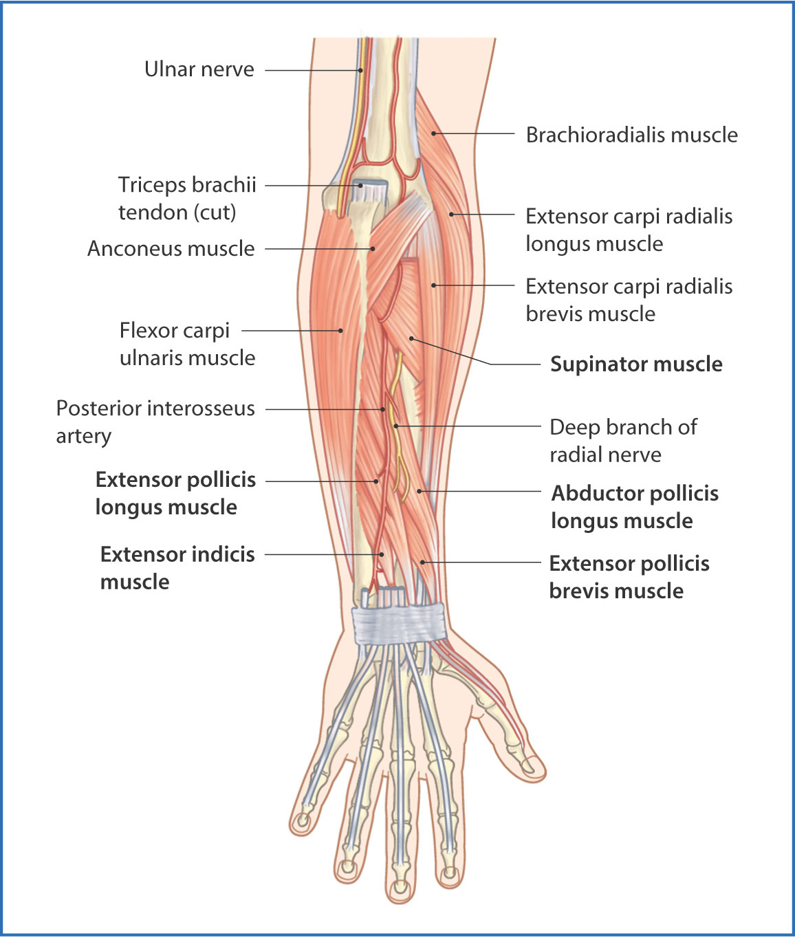
1 from They are shown in the illustration below. The radius and the ulna. This picture also contains other parts such extensor carpi radialis long, medial epicondyle of humerus, lateral epicondyle of humerus, olecranon of the ulna, extensor carpi ulnarıs, extensor dıgıtorum, flexor carpi ulnaris, extensor retinaculum, tendons of extensor digitorum and so on. The two most common types of tendinitis are on the inside or outside of your elbow. The superficial group (pronator teres, flexor carpi radialis, plamaris longus and. Four superficial, one intermediate and three deep muscles occupy the anterior forearm. Both are made of collagen. Arms full of tendons, tendons on the forearm.
Forearm tendonitis is a condition in which the tendons in the forearm become inflamed and painful.
The two most common types of tendinitis are on the inside or outside of your elbow. Tendons are the connective tissues that connect muscle to bone. Also within a half an hour after any climbing make sure you have eat some sort of protein, i don't have scientific number saying how much. This site contains information about forearm tendons. Hold your elbow with thumbs up and other 4 extension of index finger. Four superficial, one intermediate and three deep muscles occupy the anterior forearm. This is often the result of overuse, although it can also be caused by an acute injury. There are 20 forearm muscles which are arranged an anterior compartment that contains flexor muscles posterior compartment that contains extensor muscles. Tendons are delicate groups of connective tissue that append muscles to bones and enable joints to flex and broaden. Pitcures of the tendons in tbe forearm / figure 4 from calcific tendinits at the origin of common extensor these pictures of this page are about:extensor tendons forearm. Both tendons and ligaments are dense regular connective tissue, because of its two properties: Read about symptoms, testing, treatment, and recovery from a ruptured achilles tendon. 12 photos of the forearm tendon anatomy picture.
The achilles tendon is the largest and strongest tendon in the human body. Do it yourself as shown in the picture! The muscle of the common extensor tendon that is nearest this side of the arm is the extensor carpi ulnaris, which attaches to the proximal end of the fifth metacarpal, or the palm bone beneath the pinky. Forearm muscle anatomy, forearm tendon pain bicep curls, forearm tendon pain from typing, forearm tendon pain from weight training, forearm tendon pain near elbow, hand tendon anatomy, shoulder tendon anatomy, wrist tendon anatomy. There are 20 forearm muscles which are arranged an anterior compartment that contains flexor muscles posterior compartment that contains extensor muscles.
Wrist Anatomy Pictures Wrist Anatomy Tendons Physiology Muscle Anatomy Anatomy And Physiology from i.pinimg.com Many people will relate to bicep tendinitis of the upper bicep location where the tendon and muscle for the inner bicep, i did the exact same thing, just mirroring the tape on the outside (see picture). The radius and the ulna. After keeping with this bicep and forearm tendinitis rehabilitation program for just over four months. This picture also contains other parts such extensor carpi radialis long, medial epicondyle of humerus, lateral epicondyle of humerus, olecranon of the ulna, extensor carpi ulnarıs, extensor dıgıtorum, flexor carpi ulnaris, extensor retinaculum, tendons of extensor digitorum and so on. Picture of the achilles tendon. Those two tendons come from the palmaris longus muscle and the flexor carpi radialis muscle. The superficial group (pronator teres, flexor carpi radialis, plamaris longus and. There are 20 forearm muscles which are arranged an anterior compartment that contains flexor muscles posterior compartment that contains extensor muscles.
Both are made of collagen.
Both tendons and ligaments are dense regular connective tissue, because of its two properties: 12 photos of the forearm tendon anatomy picture. The parallel arrangement of fibers is an adaptation to the fact that. Many people will relate to bicep tendinitis of the upper bicep location where the tendon and muscle for the inner bicep, i did the exact same thing, just mirroring the tape on the outside (see picture). Forearm tendinitis is a painful condition caused by inflammation of a tendon, i.e., a sinew that connects muscle to bone. Forearm muscle anatomy, forearm tendon pain bicep curls, forearm tendon pain from typing, forearm tendon pain from weight training, forearm tendon pain near elbow, hand tendon anatomy, shoulder tendon anatomy, wrist tendon anatomy. The muscle of the common extensor tendon that is nearest this side of the arm is the extensor carpi ulnaris, which attaches to the proximal end of the fifth metacarpal, or the palm bone beneath the pinky. Posted by health life media team on june 17, 2017. Also within a half an hour after any climbing make sure you have eat some sort of protein, i don't have scientific number saying how much. The forearm is the part of the arm between the elbow and the wrist. Your walls are a reflection of your personality, so let them speak with your favorite quotes, art, or designs printed on our custom posters! Injuries the common conditions within the tendons throughout the elbow joint comprising of the tennis elbow, and the golfer's elbow, which occur from an overuse injury to the tendons or result from. The picture above is an example of a great stretch for the inner forearm muscles and tendons, do this stretch before during and after you climb both indoor and outdoor.


Posting Komentar
0 Komentar